Have you ever wondered what’s happening inside the mind of someone with autism when the world feels like a cacophony of overwhelming sensations? While autism is often described through behavioral observations, modern neuroscience is peeling back the layers of the brain to reveal its inner workings. Tools like functional magnetic resonance imaging (fMRI) and electroencephalography (EEG) are not just scientific marvels—they are windows into the neural symphony that shapes perception, cognition, and emotion in autism. But what do these imaging techniques truly uncover, and why do they matter in understanding this complex neurodevelopmental condition?
Imagine standing in a bustling marketplace, where every sound, color, and scent vies for your attention. For many individuals with autism, this isn’t just an analogy—it’s their daily reality. The brain’s ability to filter and prioritize sensory input is often disrupted, leading to heightened sensitivity or, conversely, a blunted response to stimuli. Brain imaging technologies like fMRI and EEG offer a glimpse into these neural mechanisms, revealing patterns that could redefine how we approach autism. But the journey to understanding these patterns is as intricate as the brain itself.
The Neural Choreography: How fMRI Maps Brain Activity in Autism
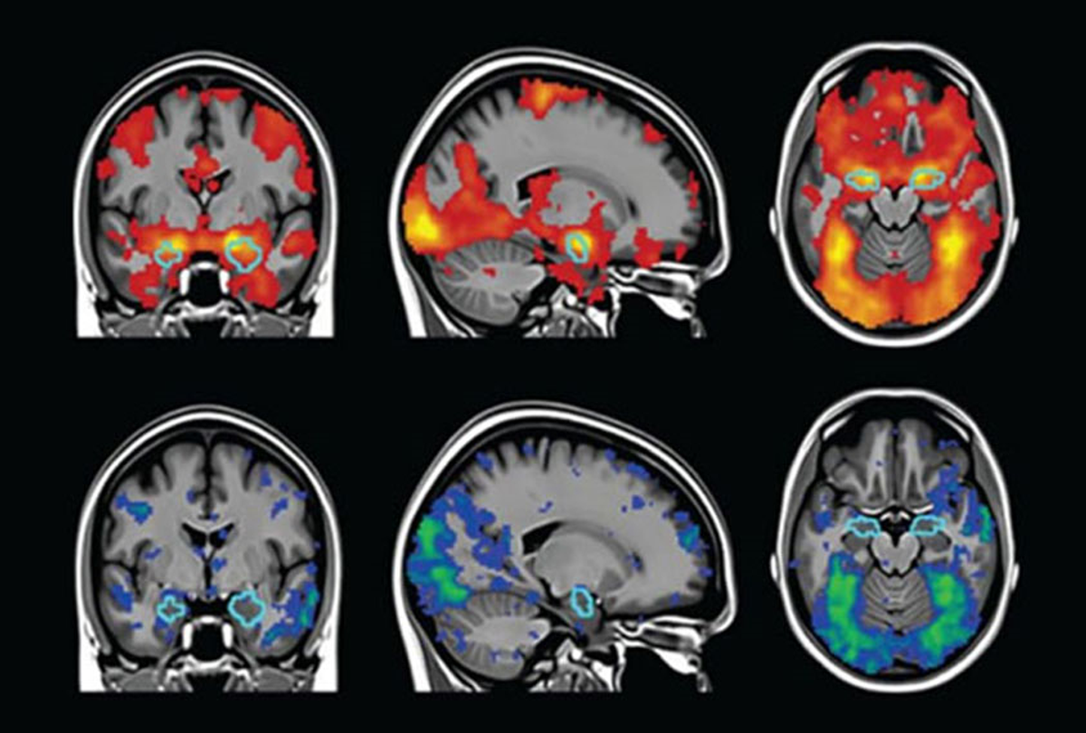
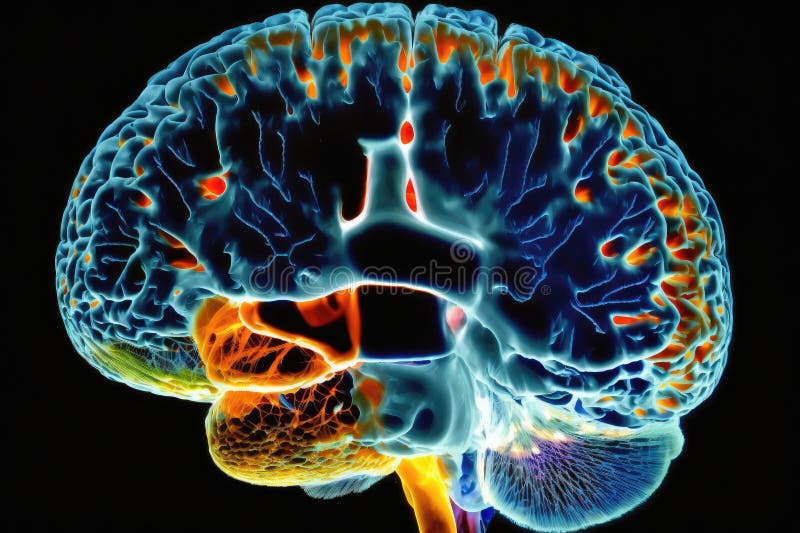
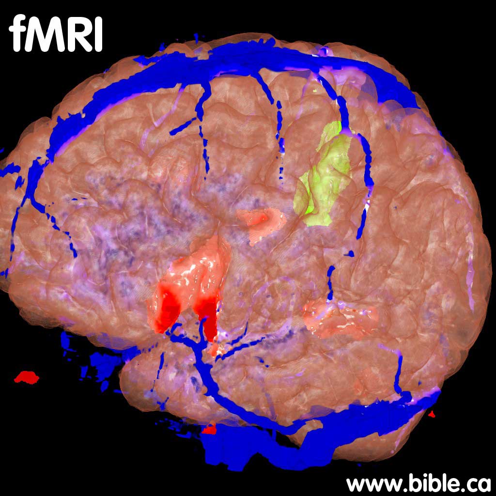
Functional MRI, or fMRI, is like a high-resolution dance recital for the brain, capturing the ebb and flow of neural activity in real time. Unlike static images from traditional MRI scans, fMRI measures changes in blood flow, which indirectly reflect brain activity. In autism research, fMRI has become an indispensable tool for identifying atypical neural connectivity and regional brain function. But what does this mean in practice?
Studies have shown that individuals with autism often exhibit hyperconnectivity in certain brain regions while displaying hypoconnectivity in others. For instance, the default mode network—a system active during rest and self-referential thought—may function differently in autistic individuals, contributing to challenges in social cognition and introspection. Meanwhile, sensory processing areas like the occipital and parietal lobes may show heightened activity in response to stimuli, explaining the sensory sensitivities commonly reported in autism.
Yet, the story doesn’t end with connectivity. fMRI has also illuminated differences in the way autistic brains process faces, a critical aspect of social interaction. The fusiform face area, a region specialized for facial recognition, often shows reduced activation in autistic individuals. This could explain why some people with autism struggle to interpret facial expressions or maintain eye contact. But is this a deficit, or simply a different way of processing the world? The answer may lie in the nuanced interplay between neural pathways and environmental demands.
/cdn.vox-cdn.com/uploads/chorus_asset/file/14368269/1j.1419979658.jpg)
EEG: The Brain’s Electrical Symphony and Its Discordant Notes in Autism
While fMRI paints a broad stroke of brain activity, electroencephalography (EEG) zooms in on the brain’s electrical whispers. EEG records the rhythmic patterns of neural oscillations, which are like the brain’s heartbeat, synchronizing communication between different regions. In autism, these oscillations often deviate from the norm, creating a symphony that’s out of tune.
One of the most striking findings in EEG studies is the prevalence of atypical gamma and beta wave activity in autistic individuals. Gamma waves, associated with attention and sensory processing, are frequently elevated, suggesting a brain that’s perpetually on high alert. This could explain the sensory overload many autistic people experience. Meanwhile, beta waves, linked to active thinking and problem-solving, may show reduced coherence, hinting at difficulties in integrating information across brain regions.
But here’s where it gets fascinating: some researchers propose that these electrical anomalies aren’t just glitches—they might be adaptations. For example, heightened gamma activity could reflect a brain that’s hyper-attuned to details, a trait that, while challenging in noisy environments, might also foster exceptional pattern recognition or memory skills. The question then becomes: Are these neural patterns deficits, or are they the brain’s way of compensating for other differences?
EEG has also shed light on the concept of “neural noise”—random, background electrical activity that can interfere with signal processing. In autism, neural noise is often elevated, which may contribute to the difficulty in filtering out irrelevant stimuli. Imagine trying to have a conversation in a room where every conversation around you is equally loud. For someone with autism, this isn’t an analogy; it’s a daily challenge. Understanding neural noise could unlock new strategies for improving focus and reducing sensory distractions in autistic individuals.
The Great Divide: Connectivity vs. Specialization in Autistic Brains
One of the most debated topics in autism neuroscience is whether the condition stems from a lack of connectivity between brain regions or an over-specialization of certain areas. The truth, as is often the case, may lie somewhere in between.
Some theories suggest that autistic brains exhibit a “local overconnectivity” phenomenon, where nearby brain regions communicate excessively, while long-range connections—those that integrate information across distant areas—are weaker. This could explain why autistic individuals might excel in tasks requiring detailed focus, such as pattern recognition or memorization, but struggle with tasks that demand holistic integration, like social navigation or multitasking.
On the other hand, other research points to a “diminished specialization” model, where certain brain regions fail to develop their typical functional roles. For instance, the amygdala, a region critical for emotional processing, may not activate as robustly in response to social stimuli in autistic individuals. This could contribute to challenges in recognizing emotions or understanding social cues. Yet, this same region might show heightened activity in response to non-social stimuli, such as objects or sensory inputs, suggesting a shift in priority rather than a deficit.
The tension between connectivity and specialization highlights a fundamental question: Is autism a story of too much focus or too little integration? The answer may depend on the individual, the task at hand, and the unique wiring of their brain. What’s clear is that the autistic brain doesn’t operate on a single spectrum—it’s a mosaic of strengths and challenges, each piece contributing to a larger, often misunderstood picture.
From Imaging to Intervention: Can Brain Scans Guide Therapy?
The insights gleaned from fMRI and EEG aren’t just academic—they hold the potential to revolutionize how we support autistic individuals. By identifying specific neural patterns, researchers and clinicians are exploring targeted interventions that could enhance quality of life.
For example, neurofeedback—a technique that trains individuals to regulate their brain activity—has shown promise in helping autistic individuals modulate their sensory responses. By using real-time EEG data, individuals can learn to adjust their neural oscillations, potentially reducing sensory overload and improving focus. Similarly, transcranial magnetic stimulation (TMS), a non-invasive procedure that uses magnetic fields to stimulate brain activity, is being investigated as a way to enhance connectivity in regions that show hypoactivity.
But the journey from imaging to intervention is fraught with challenges. Brain plasticity—the brain’s ability to reorganize itself—varies widely among individuals, and what works for one person may not work for another. Additionally, ethical considerations loom large. Could brain imaging lead to a one-size-fits-all approach to autism, ignoring the rich diversity of experiences? Or could it empower autistic individuals by providing personalized insights into their unique strengths and needs?
The key lies in collaboration. Autistic individuals, families, researchers, and clinicians must work together to translate neural data into meaningful, respectful, and effective interventions. It’s not about “fixing” the autistic brain but about understanding it—and in doing so, creating a world that accommodates its nuances.
The Unanswered Questions: What’s Next in Autism Neuroscience?
Despite the strides made in brain imaging, autism neuroscience is still in its infancy. Many questions remain unanswered, and the path forward is as complex as the condition itself.
One of the most pressing challenges is the heterogeneity of autism. No two autistic brains are alike, and what holds true for one individual may not apply to another. This makes it difficult to draw broad conclusions from imaging studies. Researchers are now turning to machine learning and big data to identify subgroups within the autism spectrum, hoping to uncover patterns that could lead to more tailored interventions.
Another frontier is the study of brain development over time. Most imaging studies focus on adults or older children, leaving a critical gap in our understanding of how autistic brains evolve from infancy through adolescence. Longitudinal studies, which track brain changes over years, could reveal when and how neural differences emerge, offering clues for early intervention.
Finally, the intersection of biology and environment cannot be ignored. How do societal attitudes, educational approaches, and sensory environments shape the autistic brain? Could a more accommodating world reduce the neural challenges associated with autism, or are these differences hardwired from birth?
The answers to these questions won’t come easily, but they’re worth pursuing. For every autistic individual who has ever felt misunderstood, every family searching for answers, and every clinician striving to make a difference, the quest to unravel the mysteries of the autistic brain is a journey worth taking.
As we stand on the precipice of discovery, one thing is clear: the autistic brain is not a puzzle to be solved but a masterpiece to be understood. With tools like fMRI and EEG lighting the way, we’re not just peering into the mind—we’re learning to listen to its unique rhythm.